Lateral cephalogram
Lateral cephalogram
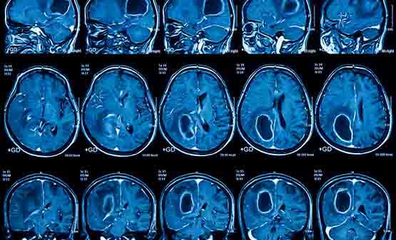
The lateral cephalogram is a fundamental diagnostic tool in dentistry and craniofacial medicine that is used to evaluate dental structures, facial proportions, and skeletal linkages. This imaging modality is essential to helping clinicians, researchers, and educators at Purdue Radiology comprehend craniofacial anatomy and disorders. Let’s examine lateral cephalograms in Purdue Radiology in more detail.
Comprehending Lateral Cephalograms: A lateral cephalogram is a two-dimensional radiographic picture that shows the side of the head and includes the teeth, jawbones, soft tissues, and skull. It helps with the diagnosis and treatment planning of a variety of orthodontic, orthognathic, and maxillofacial disorders by offering insightful information on the growth and development of facial features.
Use in Orthodontics: One of the main fields in which lateral cephalograms are used is orthodontics. These pictures are used by orthodontists to evaluate the interactions between the teeth and skeleton, spot malocclusions, and develop treatment plans. Orthodontic procedures like braces or aligners can be planned and skeletal discrepancies can be diagnosed with the use of measurements such as the ANB angle, SNA angle, and SNB angle obtained from lateral cephalograms.
Evaluation of Craniofacial Growth and Development: Purdue Radiology tracks the development of young patients’ craniofacial structure using lateral cephalograms. Clinicians can evaluate the course of skeletal maturation, forecast future growth patterns, and foresee probable orthodontic or surgical interventions by comparing sequential cephalometric photographs throughout time.
Surgical Treatment Planning: Lateral cephalograms are essential for treatment planning when orthognathic surgery or maxillofacial reconstruction is required. These pictures help surgeons plan jaw surgery movements, assess the severity of skeletal abnormalities, and forecast surgical outcomes. After surgical intervention, functional occlusion and ideal face aesthetics can be achieved with the use of accurate cephalometric analysis.
Research and Education: Lateral cephalograms are a priceless tool for Purdue Radiology research and instruction, even outside of clinical practice. The etiology of craniofacial abnormalities, biomechanics, and craniofacial morphometrics are all studied by researchers using cephalometric data. These pictures also play a crucial role in dental and medical education by giving students a fundamental understanding of craniofacial anatomy and pathology.
In conclusion, the lateral cephalogram is shown to be a flexible instrument in Purdue Radiology, with a wide range of uses from craniofacial research to orthodontic treatment planning. It is essential for the diagnosis, management, and comprehension of a wide range of craniofacial disorders due to its capacity to offer in-depth insights on the relationships between the skeleton and facial morphology. Lateral cephalograms continue to improve patient care and progress the area of craniofacial radiology due to continuous developments in imaging technology and analysis methods.
 1105/5 SADHANA 1ST FLOOR, HARE KRISHNA MANDIR ROAD, MODEL COLONY, PUNE, INDIA – 411016.
  020 2951 7802, 7744962345,
  info@purdesradiology.com